What is medical image annotation?
Medical image annotation is the process of labeling medical images to train machine learning algorithms for medical image analysis. The datasets train the model to identify various conditions or diseases in images it will encounter when it’s deployed in a healthcare setting.
Medical annotation is precisely tailored to the level of accuracy required for the best patient outcomes. Training requires large numbers of annotated images so that the model can learn typical and atypical presentations of diseases.
What kinds of medical images and documents are annotated for training datasets?
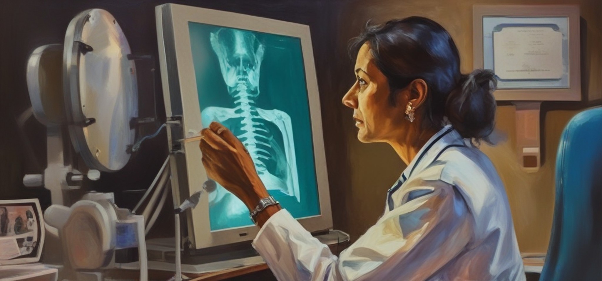
The type of image that the model will encounter in real-world use must be annotated for training, validation, and testing datasets. Annotators can label images, including X-rays, ultrasound images, and MRI and CT scans. Models can also be trained to recognize typical and atypical neuroimaging informatics technology initiative (NIFTI) scans, mammograms, electroencephalograms (EEGs), echocardiograms, and other specialized images.
AI models can also be trained to recognize medical conditions in these images, photographs or videos, as well as use audio or text data from notes to assist with diagnoses.
How does medical image annotation differ from other types of image annotation?
Because images used in healthcare diagnoses differ from other types of images, annotation processes must also differ from other image annotation techniques.
For instance, medical images may include objects in front or behind others and both solid and transparent objects. Annotators must be able to use medical image segmentation to extract “regions of interest” (ROIs) from 3D images and communicate to the AI model what each image represents. Moreover, a series may include both 2D and 3D images requiring an annotation protocol that helps train the model to recognize objects imaged using these methods in the real world.
Medical images often include several views, which requires annotating them so that the model understands they depict the same object.
Additionally, medical image annotation often focuses on very small elements or anomalies, necessitating capabilities such as zoom and contrast enhancement, which may not be necessary in other types of annotations.
How to prepare data for medical image annotation
Before a data annotator can label an image to train an AI model to identify objects or patterns in a medical scan, images must be formatted correctly, and teams must ensure they comply with regulations throughout the process.
Image format
Medical images are in digital imaging and communications in medicine (DICOM) format, unlike other images that may be in typical image formats such as JPEG or PNG.
Medical image software also generates images that typically are high resolution. For example, mammogram images can be up to 5 million pixels, and files can be several gigabytes. Annotators need tools that support the size of the images and enable labeling them.
Compliance
Anyone handling or viewing medical images must follow Health Insurance Portability and Accountability Act (HIPAA) regulations. Additionally, all images prepared for annotation should be deidentified and protected by data processing agreements.
Challenges with medical image annotation
Any image annotation project requires creating a strategy that will result in a model that produces optimal outputs. For example, AI project teams need to determine the best annotation method (e.g., image classification, object detection, or segmentation) and annotation shapes to use within images. However, medical image annotation requires taking additional factors into account to ensure a successful project, including:
Data preparation time
Images chosen for the dataset must be cleaned, formatted, and carefully selected. Images that don’t provide valuable information to the model should be discarded from the set to ensure the platform delivers accurate results.
Planning for data preparation time will ensure annotators don’t have to choose between quality and making deadlines when completing their work.
Finding skilled annotators
Medical image annotation requires data labelers with annotation and healthcare expertise. Medical image annotators must be skilled in dealing with transparencies and work with detailed precision. They also need to annotate multiple views so that the model understands they all represent the same object.
Achieving accuracy
Labeling consistency is critical. If annotators use different techniques to label images, the model will work with less accuracy than if all annotators had all followed the same protocol. A quality control process is crucial to ensure image annotation accuracy and, ultimately, the best AI model performance.
Limited access to data
Properly training an AI model requires a dataset that includes a large number of images. However, the dataset must represent all types of images the model will encounter in the real world.
Human physiology can vary based on race, geographic region, and other factors – and data from all areas may not be readily available. If the model is trained on only particular images rather than any it could encounter, it could introduce bias into the results.
Data bias is unacceptable
If an AI model is biased, the result can literally be fatal. If the model is trained only with images from a portion of the population, anomalies in images from other groups may go undetected. Diagnoses may be missed at a higher rate for specific demographics, which can lead to delays in treatment and negative outcomes.
Patient privacy
Healthcare providers and their AI project teams may have strong policies in place to protect patient privacy. Those policies must extend to or align with data annotators preparing training datasets.
The benefits of medical image annotation
When a fully trained machine learning model automates image interpretation, patients don’t have to wait hours or even days for a radiologist to review images. When image interpretation is used as an aid of diagnostics doctors can review the interpretation with greater confidence in what they’re looking for. The aid of diagnostics also reduces the liability of medical professionals, as they can trust that a well-trained algorithm has already taken a preliminary review of the results, and they can review the interpretation with greater confidence in what they’re looking for.
Additionally, precise medical image annotation enables AI to detect anomalies that may not be visible to a physical reviewing of the image. AI can also detect biomarkers early in disease progression that may not be visible to the human eye. Although annotation techniques that enable this capability are still developing, this is an important advancement, albeit still in its in
fancy, that will enable early diagnoses.
A study at Stanford found that an AI algorithm matched experts in accuracy and decreased the time to read an image from 4 hours to less than 2 minutes for chest X-rays taken to identify 14 different conditions.
Additionally, as radiology and other medical fields face labor shortages, AI platforms trained with datasets prepared with precise medical image annotation can help fill gaps and enable healthcare organizations to provide care to more patients.
Medical image annotation not only trains models that enhance patient care. It can also help create models that accelerate medical research and recognize patterns in massive datasets that may go unnoticed using traditional processes.
What specific fields utilize it most?
Virtually any area of medicine that uses imagery can benefit from AI, including these specialties that are benefitting from AI today:
Oncology
Early detection is key to cancer patients’ survival. Medical images with precisely annotated images of early stages of disease progression increase the chances of good patient outcomes. A Tulane University study found that AI can detect colorectal cancer in tissue samples as well as or better than pathologists.
Neurology
Medical image annotation can be used to train a machine learning model to recognize brain tumors, blood clots, injuries, and neurological disorders that are revealed in CT or MRI scans. Additionally, AI is helping researchers understand how changes in the brain manifest, how to obtain insights about mental disorders, and possibly how to discover effective treatments.
Dentistry
AI can quickly and accurately detect dental issues, including gum disease, compromised tooth structure, and decay. AI can also assist with orthodontic treatment planning and diagnosing osteoarthritis in the temporomandibular joint.
Hepatology
Diagnosing liver, gallbladder, bile ducts, and pancreas diseases and conditions can be challenging. Traditionally, diagnoses depended on practitioners evaluating images based on their training and experience. Unfortunately, bias can impact a physician’s interpretation and possibly lead to a missed diagnosis.
Machine learning models trained on thousands of properly annotated images based on known conditions can assess images without bias, increasing overall accuracy.
Ophthalmology
Images can reveal retinal diseases, such as diabetic retinopathy and macular degeneration, and conditions, such as retinal tears or detachment. AI can provide highly accurate and detailed results within minutes.
Dermatology
AI models can learn to identify skin conditions from images. This saves time, enables faster treatments, and could help specialists to care for more patients via telehealth.
What does the future of medical annotation look like?
The success that AI has had with medical imaging will result in more adoption and more use cases, which will lead to a greater demand for medical image annotation. Furthermore, medical images make up around 90% of healthcare data. To enable AI platforms to use this data to help doctors with future diagnoses, it must be expertly labeled for specific use cases.
Achieving highly accurate medical annotation at scale is often difficult for in-house teams. Partnering with an experienced medical image annotation service is the best way to ensure accuracy, speed, and quality control.
When you partner with Sigma.ai, you can also leverage our experience with image selection and annotation to ensure the dataset has the right degree of variability for optimal model performance. You will also have the advantage of using our technology that predicts annotations using pre-trained models, which are then refined by humans in the loop, helping you produce an effective dataset more quickly.
The integrity of healthcare AI models rests on both precision and patient safety. Before starting your project, you must secure your data. Learn about the critical protocols and compliance standards in Ensuring data privacy and security in data annotation. To see the uncompromising level of technical quality required for image-based AI, view our case study on achieving Pixel-perfect image annotation for product recognition: A case study.
Collaborate with Sigma to power your healthcare AI with data you can trust.